 dr.vision
< 1 min
is how long the injection itself takes. With preparation and a check afterward, you are at the practice for about 1–1.5 hours.
Start here
What suits you?
Find out in 2 minutes which procedure suits your eyes.
Start the Suitability CheckOr by age:
< 45yearsGet rid of glasses & contact lenses 45–64yearsGet rid of reading glasses 65+yearsCataract & glasses-free★★★★★ 5.0 · 376 Google reviews · 20,000+ procedures
Decide well
What you should know beforehand.
Who operates?Prof. Dr. Mehdi ShajariFOCUS Top Doctor · operates on every patient personally What does it cost?Laser from €3,498 · Lenses from €6,886Prices for both eyes Is it safe?Safety & risks When will I be fit again?Work, sports, driving Would you rather read in peace first?Decision Guide Freedom from GlassesProcedures
Laser or lens?
Laser eye surgery
SMILE ProSmartSightFemto-LASIKTrans-PRKPRKPresbyond (laser away your reading glasses)All laser procedures at a glanceLaser procedures compared ›Lens surgery
ICLLens exchange (reading glasses)Cataract surgery with premium lensLens types: trifocal, EDOF, toricLens types compared ›Start here
Something wrong with your eyes?
A thorough eye check-up with us clarifies what is behind it.
Book an eye check-up★★★★★ 5.0 · 376 Google reviews
Injection therapy · IVOM
A medication exactly where it works.
In wet macular degeneration, diabetic macular edema or retinal vein occlusion, an injection into the eye can protect your retina. The injection itself takes less than a minute, and thanks to numbing drops, most people feel only brief pressure. Prof. Dr. Mehdi Shajari treats you personally.
 12× FOCUSTop Doctor
A few seconds. Big effect.
< 1 min
is how long the injection itself takes. With preparation and a check afterward, you are at the practice for about 1–1.5 hours.
hardly
Numbing drops. Most people feel only brief pressure, like a touch.
4–16 wks
Interval between two injections, depending on the medication and how the retina responds.
GOÄ
Fee and medication billed separately, cost estimate in advance.
376 reviews. Average 5.0.
“Unbeatable that you can get appointments on weekends too! I was helped in the best way, so that it almost annoys me that I struggled with my problems all those years. Can be recommended without reservation!”
“I was able to book an appointment online right away, did not have to wait until it was my turn. … Overall I am very satisfied and simply totally thrilled.”
“We had an appointment with Prof. Shajari this Friday … and had a great experience all round with the practice. … So a clear recommendation of the practice to others!”
“The most professional and best-equipped eye doctor's practice I have ever visited. I am very satisfied with the service and will continue to use the practice.”
IVOM in five answers.
For anyone with three minutes. Each answer leads to the detailed chapter.
The intravitreal operative medication injection: a medication is injected into the vitreous with a very fine needle. From there it works directly at the retina, in an amount of less than one drop.
To the chapterMainly in wet macular degeneration, diabetic macular edema, retinal vein occlusion and new blood vessel growth in strong nearsightedness. Goal: draw fluid out of the retina and preserve vision.
ConditionsHardly. The eye is numbed with drops, and the needle is thinner than for a blood draw. Many describe a brief pressure. Afterward, the eye may feel scratchy for a few hours.
Fear and painAt the start, usually three to five injections about four weeks apart, depending on the medication and the disease, then every 4 to 16 weeks depending on the findings, sometimes longer. In wet AMD, the therapy often runs for years.
Treatment schedulesdr.vision is a private practice and bills according to the GOÄ; the medication is added. Private health insurance and Beihilfe usually reimburse. Patients with statutory health insurance pay themselves with us.
CostsNo drop reaches the retina. The injection does.
In an IVOM, a medication is injected into the vitreous with a very fine needle. Eye drops do not reach the retina, and tablets only in a small amount. That is why the medication is given directly into the vitreous, the clear gel inside the eye. From there it reaches the macula, the spot of sharpest vision, and works for weeks.

VEGF
VEGF makes diseased vessels grow and become leaky. Anti-VEGF medications catch it: fluid in the retina goes down, and bleeding becomes less frequent.
Depot
In some forms of macular edema, a small cortisone implant works better or longer. It releases the active substance over months.
0,05 ml
That little is enough because the medication is right at the target. Very little of it reaches the rest of the body.
IVOM stands for intravitreal operative medication injection. “Intravitreal” means into the vitreous. It counts as “operative” because the eye is opened in the process; that is why the same hygiene rules apply as for surgery: sterile room, disinfection, sterile drape, face mask. Internationally, it is called intravitreal injection (IVI).
The injection treats the consequences, that is, fluid, swelling and vessel growth, not the cause. When the effect wears off, the fluid returns. That is why it is repeated. In vein occlusions and diabetic macular edema, the intervals can become very long over time or the treatment can end entirely; in wet AMD, it usually runs for years.
When the retina draws water.
When diseased vessels or leaky vessel walls let fluid into the macula. This happens mainly in wet age-related macular degeneration, in diabetic macular edema and after a retinal vein occlusion. In wet AMD, time counts: the earlier the first injection, the more vision is preserved. International recommendations, for example from the British professional society, call for treatment to begin within two weeks of diagnosis.
New, leaky vessels under the macula. Distorted vision, a dark spot in the center, often within weeks.
Anti-VEGF
DiabetesThe vessel walls become permeable, and the macula swells. Reading becomes difficult, and colors look paler.
Anti-VEGF, cortisone
A retinal vein is blocked, and blood and fluid back up. Often with high blood pressure.
Anti-VEGF, cortisone
With high nearsightedness, vessels can grow in under the macula.
Anti-VEGF
| Disease | What the injection does | Typical number in year 1 | |
|---|---|---|---|
| Wet (neovascular) AMD | stops vessel growth and leakage; vision stays stable in most people, some gain lines | 6–9 | Macular degeneration |
| Diabetic macular edema | reduces swelling; vision often improves | 6–9 | Diabetes & retina |
| Vein occlusion with macular edema | reduces edema, prevents new vessel growth | 6–8 | Retina examination |
| Myopic CNV | closes the new vessel | 1–3 | Retina examination |
| Macular edema with inflammation (uveitis) | cortisone implant calms the inflammation | 1–2 implants | Retina examination |
| Proliferative diabetic retinopathy | Anti-VEGF as an alternative or addition to the laser | depending on findings | Diabetes & retina |
Straight lines become wavy, a gray spot in the center, faces blur: this can be wet AMD. An appointment within a few days, not weeks.
The retinal scan shows fluid in thousandths of a millimeter. It decides whether, how often and with what an injection is given.
One principle. Several active substances.
Usually an anti-VEGF medication that blocks the growth factor VEGF. The active substances differ mainly in how long they work: from about 4 weeks to 16 weeks and more. In certain edemas, a cortisone implant is used. Which medication fits depends on the disease, the findings and how you respond.
| Active substance | Group | Approved in the EU for, among others | Interval after loading phase | Good to know |
|---|---|---|---|---|
| Ranibizumab | Anti-VEGF, antibody fragment | wet AMD, DME, vein occlusion, myopic CNV | 4–12 weeks | proven for many years, biosimilars available |
| Aflibercept 2 mg | Anti-VEGF, fusion protein | wet AMD, DME, vein occlusion, myopic CNV | 8–16 weeks | often used in a treat-and-extend schedule, biosimilars available |
| Faricimab | bispecific antibody (VEGF-A and angiopoietin-2) | wet AMD, DME, vein occlusion | up to 16 weeks | blocks two signaling pathways |
| Brolucizumab | Anti-VEGF, small antibody fragment | wet AMD, DME | up to 12–16 weeks | rarely inflammation of the retinal vessels, hence careful selection |
| Dexamethasone implant | Cortisone, depot | DME, macular edema after vein occlusion, uveitis | 3–6 months | can raise eye pressure and speed up a cataract |
| Fluocinolone acetonide implant | Cortisone, long-term depot | chronic DME with insufficient response, uveitis | up to 3 years | for eyes that do not respond enough to other therapies |
We deliberately name only active substances, not brand names. Approvals according to the European Medicines Agency (EMA, EPAR) and prescribing information; intervals are typical ranges.
When the patent protection of a biological medication expires, other manufacturers may offer very similar products. Such biosimilars exist for ranibizumab and aflibercept 2 mg. They must show in studies that they work and are as safe as the original. Whether a biosimilar comes into question for you, we discuss with you, also with a view to the costs.
If fluid stays in the retina despite regular injections or the intervals have to stay very short, a switch to an active substance that works longer or differently can help. We decide this based on the OCT images over time, not by feel.
Ten minutes in the procedure room.
You are awake, lying comfortably, and the eye is numbed with drops. After thorough disinfection, the medication is given into the vitreous from the side with a very fine needle. The injection itself takes only a few seconds, the whole procedure about 10 minutes.
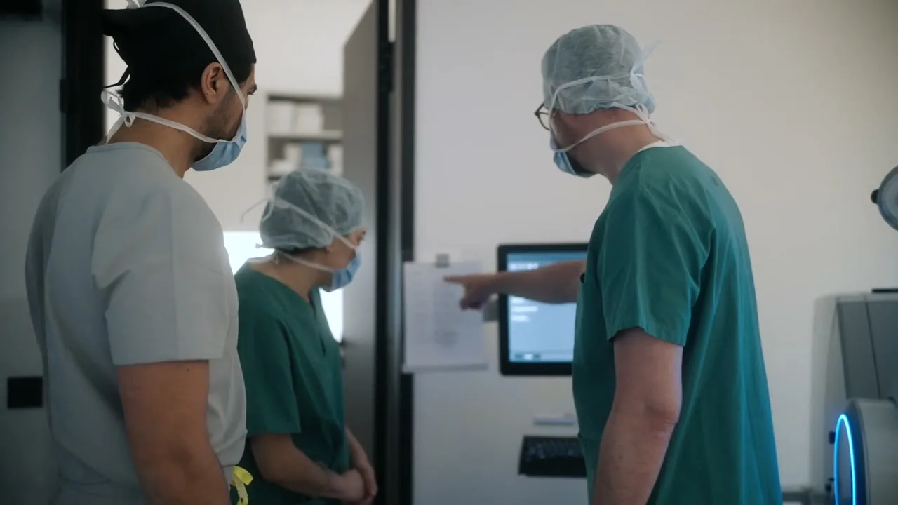

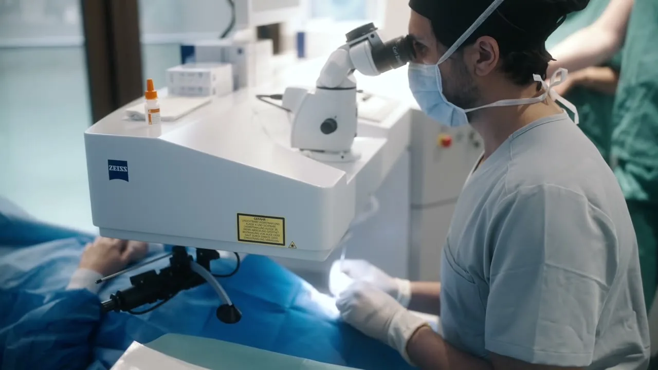
 0:00 · Arrival
You lie on a comfortable couch. We check which eye and which medication, then drops numb the surface. If you like, we tell you each step.
0:03 · DisinfectionEyelids, lashes and the surface of the eye are disinfected with an iodine solution, and the skin around them is covered. This is the most important protection against infection. Many feel the stinging more than the injection.
0:06 · Eyelid holderThis way you do not have to fight the blink reflex. The injection site is marked: from the side, 3.5–4 mm behind the edge of the cornea, far away from the lens and the center of the retina.
0:08 · InjectionYou look in one direction, the fine needle goes through the sclera, and the medication flows into the vitreous. Most people feel a brief pressure. Afterward you often briefly see streaks or a small bubble.
0:10 · CheckWe check whether you can count fingers and, if needed, your eye pressure. You will get drops and an eye patch for the way home. You must not drive yourself on this day.
Eat normally and take your medications, including blood thinners, unless agreed otherwise. No makeup around the eye. Having someone with you is nice but not necessary; you must not drive yourself on this day.
Around 1–1.5 hours at the practice, of which around ten minutes in the procedure room. Before the injection, we image your macula with OCT and Clarus.
Remove the patch at home, use the drops as planned, do not rub your eyes. The day after the injection, we check the eye; if you come through your eye doctor, the follow-up check usually takes place there. Daily life is then usually back to normal.

At dr.vision
At dr.vision, Prof. Dr. Mehdi Shajari gives the injection personally. In a sterile procedure room, with OCT monitoring over the course of treatment.
Prof. Dr. Mehdi Shajari always performs the treatment for macular degeneration personally.
High-resolution OCT cross-sectional images and ultrawide retinal images without pupil dilation show when the next injection is needed.
Medication, interval and next appointment on one sheet, also for your family doctor or diabetes specialist.
As often as necessary. As rarely as possible.
At the start, there are usually three to five injections about four weeks apart. After that, the interval depends on the retina: with the treat-and-extend schedule, the next interval is extended by 2 weeks if the retina is dry and shortened again if there is new fluid, up to 16 weeks and more. This way you get no injection too many and none too late.
| Schedule | How it works | Advantage | Disadvantage |
|---|---|---|---|
| Fixed (monthly / every two months) | Injection at a fixed interval, regardless of the findings | best visual results in studies | many appointments, some injections unnecessary |
| As needed (PRN) | monthly follow-up check with OCT, injection only if there is fluid | fewer injections | many follow-up appointments; relapses are only treated once they have occurred |
| Treat-and-extend (T&E) | inject every time, extend or shorten the interval depending on the findings | good results with fewer appointments; follow-up check and injection on the same day | injection is given even when the retina is dry |
The thought is worse than the injection.
Most patients say after the first injection that they worried far too much. Numbing drops take away the pain, the needle is hair-thin, and you do not see it coming: you look in another direction. Many feel only a brief pressure of 1–2 seconds.
| When | What you feel | What helps |
|---|---|---|
| Before | Tension, racing heart, especially the first time | Ask questions beforehand, bring someone with you, breathe calmly; on request, a sedative after consultation |
| Disinfection | Burning from the iodine solution | Eyes closed, breathe calmly; it passes after seconds |
| Injection | brief pressure, no sting; some people notice nothing at all | look in the direction we announce, we count along |
| First hours | Scratchiness, foreign body sensation, tearing | Artificial tear drops, keep the eye closed, do not rub |
| First days | black dots or streaks, a red spot on the white of the eye | normal; they disappear within days to two weeks |
You look to the side or upward, and the needle comes from the outside. The light of the microscope is somewhat dazzling, but you see nothing more.
A lid speculum holds the eye open, so blinking is no problem. If you need to cough or sneeze, tell us and we wait.
If you wish, you hear beforehand what happens next. If you prefer not to hear anything, you get quiet.
From the second or third injection, you know what to expect. For most people, the fear decreases noticeably.
Tell us beforehand. We plan more time and, on request, have someone accompany you in the room.
For wet AMD and macular edema, there are currently no equally effective drops or tablets. Waiting often costs permanent vision in wet AMD.
Scratchiness yes. Increasing pain no.
An irritated eye, a red spot on the white of the eye and black dots in your field of vision are normal in the first days. Increasing pain, clearly worse vision or strong redness in the days after the injection can be an infection: in that case, call us immediately.
| What you notice | What it may mean | What you do |
|---|---|---|
| Scratchiness, tearing, foreign body sensation in the first hours | Irritation from the disinfection and lid speculum | Artificial tears, close your eyes; usually passes by evening |
| Red spot on the white of the eye | small bleed under the conjunctiva at the injection site | harmless, disappears in 1–2 weeks |
| Black dots, streaks, a bubble | Medication or a small air bubble in the vitreous | normal, disappears in days |
| Increasing pain, stronger redness, worse vision in the days after the injection | possible infection (endophthalmitis) | call immediately, treatment on the same day |
| Suddenly many new dots, flashes, a shadow like a curtain | possible retinal detachment | Call immediately |
| Severe headache and eye pain with nausea | Pressure rise | Call immediately |
| Activity | After the IVOM |
|---|---|
| Driving | not on the day of the injection; the next day if you can see well again |
| Work, screens | usually the next day |
| Showering, washing hair | from the next day, no water or shampoo in the eye |
| Eye makeup | after 2–3 days |
| Swimming, sauna | after 3–7 days |
| Sports, flying | Sports after 1–2 days; flying is allowed |
If you have questions after the injection, you can reach us 24 hours a day at 069 509 586 770.

Injection recommended and unsure?
Let us talk about it. Call us.
Jana from our Patient Care team schedules the retina examination with OCT, also as a second opinion on an ongoing injection therapy. Bring your previous findings. Call around the clock, WhatsApp Mon–Fri 8:00–19:00, Sat 9:00–15:00.
Your path. In four steps.
From the first diagnosis to routine: you know what is happening at every appointment. Diagnosis and plan are usually given at one appointment, and the first injection for wet AMD as soon as possible. International recommendations call for treatment to start within two weeks of diagnosis.
Step 1 of 4

Clarify whether the retina needs an injection.
Bring: previous findings, medication list, and with diabetes the latest HbA1c value.
Arrange an examinationStep 2 of 4

Decide on the medication and schedule.
Informed consent takes place in good time before the first injection, not only in the procedure room.
The medicationsStep 3 of 4
Ten minutes, sterile, personal.
If needed, both eyes on the same day, each with its own sterile set.
The procedure in detailStep 4 of 4
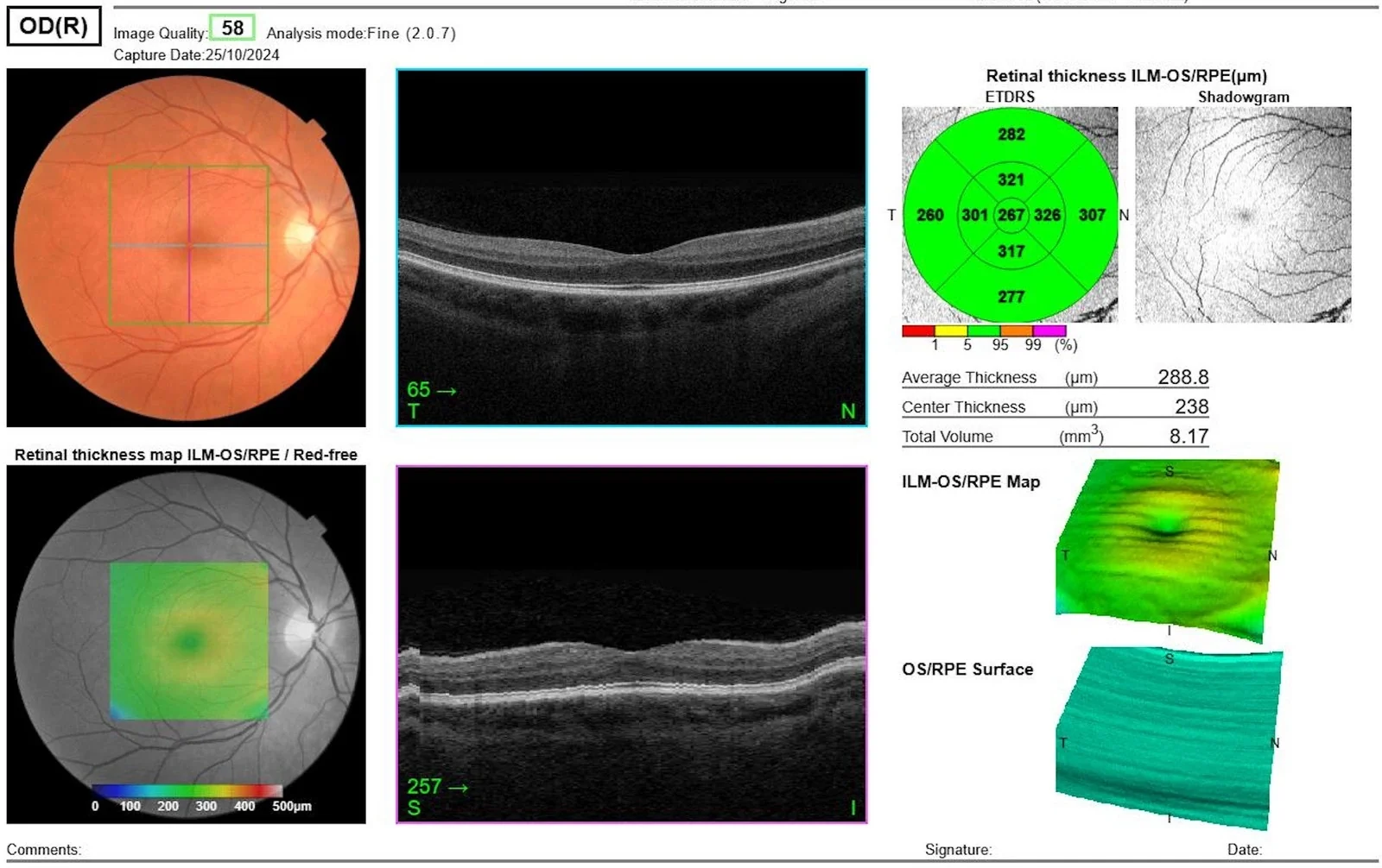
OCT decides the next interval.
Reminder for every appointment from Patient Care. Questions in between via WhatsApp.
Treatment schedulesRarely serious. Important: know the warning signs.
The most common side effects are harmless: a red spot on the white of the eye and black dots for a few days. The most dangerous complication, an infection in the eye, occurs in about 1 in 1,800–2,000 injections and can be treated if it is detected early.
A small, sharply defined red spot on the white of the eye at the injection site. Does not hurt.
about 1 in 10
more common with blood thinners.
Nothing necessary, it disappears in 1–2 weeks. Blood thinners are not stopped.
The additional volume raises the pressure for minutes. With cortisone implants, it can stay raised for weeks.
short-term almost always; lasting with cortisone about 1 in 3
Follow-up check after the injection, especially with glaucoma; with cortisone, regular pressure measurement and drops if needed.
Germs get into the inside of the eye. Signs usually appear in the first days, rarely only after a week: increasing pain, redness, worse vision.
about 1 in 1,800–2,000 injections
Iodine disinfection, sterile technique, face mask; clear warning signs, emergency number, treatment on the same day.
The needle or vitreous traction injures the retina. Signs: flashes, many new dots, shadows.
under 1 in 5,000
Injection site far forward at a safe distance; if there are warning signs, immediate examination, laser or surgery.
A reaction to the medication, usually mild. With one active ingredient (brolucizumab), rarely with inflammation of the retinal vessels.
rare, depending on the active ingredient
Careful choice of active ingredient, follow-up checks, anti-inflammatory drops.
Cortisone implants accelerate a cataract.
common with repeated implants
Use with restraint in eyes with their own lens; a cataract can be operated on well, see cataract.
Anti-VEGF reaches the blood in tiny amounts. A connection with heart attack or stroke has not been clearly shown in large studies.
very rare
Ask about previous illnesses; after a recent heart attack or stroke, weigh benefit and risk especially carefully.
Frequencies from specialist literature and prescribing information.

Because it concerns your central vision.
A surgeon who injects personally, retinal diagnostics that show the smallest changes, and a team that accompanies you over the years. 5.0 stars from 376 reviews.
Prof. Dr. Mehdi Shajari. Personally.
Prof. Dr. Mehdi Shajari is a specialist physician and professor of ophthalmology (German title: außerplanmäßiger Professor) and was a senior physician (Oberarzt) at the university hospitals of Munich and Frankfurt. He performs the injection therapy for macular degeneration personally. Senior physician (Oberärztin) Ivana Pavlovic and Dr. Claudia Grunwald assess your retina and draw up the treatment plan with him. You are looked after not by changing doctors, but by the same team.
What you get with us.
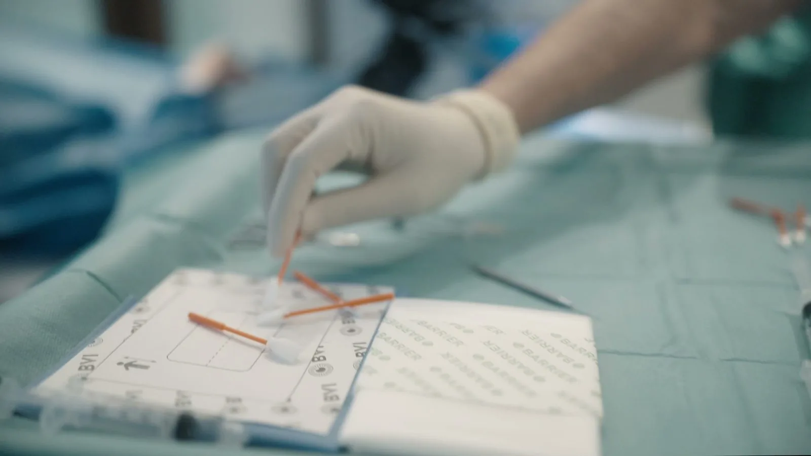
One team
Prof. Dr. Mehdi Shajari injects personally.
Reviews
5,0
376 Google reviews
Retina
The interval follows the findings, not the calendar.
OCT cross-sectional images and ultrawide retinal images, usually without pupil dilation. This is how we decide on every injection based on measurable findings.
5,0
★★★★★
from 376 Google reviews
Whoever knows your retina sees changes sooner.
Surgery
Own procedure room, sterile draping, iodine disinfection.
As few injections as possible, as many as necessary.
Medication, schedule, fear: we take the time until everything is clear.
Follow-up check and injection usually at the same appointment.
Billing per the German fee schedule for physicians (GOÄ), written cost estimate before every treatment.
Jana answers questions about appointments, findings and costs via WhatsApp, usually within 10–15 minutes, Mon–Fri 8:00–19:00, Sat 9:00–15:00. We are available by phone around the clock.
Patient Care
Junghofstraße 16, free parking in the building.
A look inside the practice and the team.




Two items: treatment and medication.
An IVOM consists of the medical fee under the German fee schedule for physicians (GOÄ) (procedure, examinations, OCT) and the medication, whose price varies greatly depending on the active ingredient. Private health insurance and Beihilfe (allowance for German civil servants) usually reimburse medically necessary therapy. dr.vision is a private practice; patients with statutory health insurance pay themselves with us, while in practices contracted with statutory insurers, the IVOM is a covered service for an approved indication.
According to the GOÄFee and medication billed separately · written cost estimate in advance
With an approved indication, the IVOM is medically necessary; private health insurers reimburse the fee and medication depending on the plan, and an out-of-pocket share may remain. For expensive active ingredients, some insurers ask beforehand. You receive a cost estimate in advance.
The medically necessary services under GOÄ and the medication are eligible for Beihilfe. Your private supplementary insurance reimburses the remaining share depending on the plan; an out-of-pocket share is possible. We word the cost estimate accordingly.
dr.vision has no statutory health insurance license; you pay the fee and the medication yourself. You can declare costs you bear yourself as extraordinary expenses in your tax return. Whether and how much the tax office recognizes depends, among other things, on your reasonable burden, see tax.
| Treatment | Intravitreal surgical drug administration: injection into the vitreous |
|---|---|
| Conditions | wet AMD, diabetic macular edema, vein occlusion, myopic CNV, macular edema in uveitis |
| Medications | Anti-VEGF (ranibizumab, aflibercept, faricimab, brolucizumab), cortisone implants |
| Process | Numbing drops, disinfection, injection in a few seconds; around 10 minutes in the procedure room |
| Pain | usually only brief pressure |
| Schedule | Start usually with 3 injections 4 weeks apart, then 4–16 weeks depending on the findings (treat-and-extend) |
| Afterward | do not drive yourself on the day of the injection; daily life usually from the next day |
| Most common serious risk | Infection in about 1 in 1,800–2,000 injections |
| Costs | Fee under GOÄ plus medication |
| Insurance | private health insurance and Beihilfe under GOÄ; patients with statutory health insurance pay themselves at dr.vision |
| Surgeon | Prof. Dr. Mehdi Shajari |
| Location | Junghofstraße 16, 60311 Frankfurt am Main |
Go deeper.
This page answers the most important questions. For related topics there are separate pages, each with context and the questions from consultation hours:
Dry or wet, Amsler test, nutritional supplements and when injections are given.
To the AMD pageDiabetesFollow-up intervals, stages, macular edema and laser or injection.
Learn morePreventive careRetina, lens, eye pressure: finding early changes before they cause trouble.
To the check-upLensWhen the lens becomes cloudy after cortisone implants or with age.
To the cataract pageVitreousBlack dots in your field of vision: when harmless, when an emergency.
Learn moreSecond opinionWhen the injections do not work or are needed very often.
Get a second opinionStill have questions?
The questions we hear most often in consultation hours, with our answers. Prof. Dr. Mehdi Shajari
Hardly. The eye is numbed with drops beforehand, and the needle is very thin. Most people feel only a brief pressure of one to two seconds. Many find the burning of the disinfection more unpleasant. Afterward, the eye may feel scratchy for a few hours.
No. You look in a direction we announce, and the needle comes from the side. At most you see the bright light and then briefly streaks or a small bubble when the medication flows into the vitreous.
The injection itself takes a few seconds, the procedure with disinfection around ten minutes. With check-in, examination and the follow-up check afterward, you should plan on about one to one and a half hours.
No, as a rule not. Blood thinners only increase the likelihood of a harmless red spot on the white of the eye. Do not stop your medications on your own; but do tell us which ones you take.
Yes, if both eyes need treatment. Each eye is then treated like a separate procedure, with its own disinfection and its own sterile set. This saves you appointments.
Not on the day of the injection. The eye is irritated, and vision is limited by drops and streaks. Bring someone with you or use bus, train or taxi. The next day you may drive if you can see well again.
Increasing pain, growing redness or clearly worse vision in the days after the injection can be an infection. Flashes, many new dots or a shadow like a curtain are also a warning sign. In that case, call immediately, on the same day.
At the start, usually three, four weeks apart. After that, it depends on the retina: with wet AMD, it is often six to nine in the first year, fewer later. With diabetic macular edema and after vein occlusions, the intervals can become very long over time or the therapy ends.
With wet AMD, the fluid usually returns, and with every relapse, vision can be lost permanently. Talk to us before you end a therapy: often the interval can be extended or the medication changed instead of stopping altogether.
The main goal is to preserve vision. Many patients additionally regain visual acuity, especially if treated early and no scar has yet formed in the macula. With diabetic macular edema, improvement is common.
For wet AMD and macular edema, injections are currently the most effective treatment. Lasers play a role in diabetic macular edema and proliferative retinopathy; drops or tablets do not replace the injection.
The costs are made up of the medical fee under GOÄ and the medication; they differ considerably depending on the active ingredient. You receive a written cost estimate before starting. Private insurers and Beihilfe usually reimburse; patients with statutory health insurance pay themselves with us.
In practices and clinics accredited for statutory insurance, yes, for an approved indication. dr.vision is a private practice without statutory insurance accreditation; patients with statutory health insurance pay for treatment with us themselves. Many use us for diagnosis, a second opinion or when they want treatment by a particular doctor.
Medically reviewed by Prof. Dr. Mehdi Shajari · As of October 2026
Everything important to read again and pass on.
Arrives by e-mail in about 60 seconds. No newsletter, just the guide and, if you wish, a follow-up from Patient Care. Privacy policy
How would you like to continue?
Online, by phone at 069 509 586 770 (available 24 hours) or via WhatsApp. The first appointment is a retina examination: visual acuity, OCT cross-section scan of the macula, retinal image and a talk about whether and with what an injection should be given. You go home with a written treatment plan.
In Frankfurt, Junghofstraße 16 · OCT included
Our locations.
 Opening hours
Junghofstraße 16, 60311 Frankfurt am Main
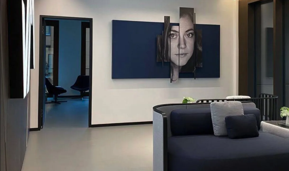 Opening 2027
Breite Straße 69, 40213 Düsseldorf (in the Neuer Stahlhof, corner of Graf-Adolf-Platz)
Until opening: preliminary talk in Frankfurt, Patient Care plans your travel.
Plan route Opening 2027
Lautenschlagerstraße 23, 70173 Stuttgart (2nd floor, step-free access)
Until opening: preliminary talk in Frankfurt, Patient Care plans your travel.
Plan route