 dr.vision
1–2 years
without retinal changes, depending on general risk. More often with changes.
Start here
What suits you?
Find out in 2 minutes which procedure suits your eyes.
Start the Suitability CheckOr by age:
< 45yearsGet rid of glasses & contact lenses 45–64yearsGet rid of reading glasses 65+yearsCataract & glasses-free★★★★★ 5.0 · 376 Google reviews · 20,000+ procedures
Decide well
What you should know beforehand.
Who operates?Prof. Dr. Mehdi ShajariFOCUS Top Doctor · operates on every patient personally What does it cost?Laser from €3,498 · Lenses from €6,886Prices for both eyes Is it safe?Safety & risks When will I be fit again?Work, sports, driving Would you rather read in peace first?Decision Guide Freedom from GlassesProcedures
Laser or lens?
Laser eye surgery
SMILE ProSmartSightFemto-LASIKTrans-PRKPRKPresbyond (laser away your reading glasses)All laser procedures at a glanceLaser procedures compared ›Lens surgery
ICLLens exchange (reading glasses)Cataract surgery with premium lensLens types: trifocal, EDOF, toricLens types compared ›Start here
Something wrong with your eyes?
A thorough eye check-up with us clarifies what is behind it.
Book an eye check-up★★★★★ 5.0 · 376 Google reviews
Diabetes & retina · Diabetic retinopathy
Look every one to two years. So that it stays that way.
With diabetes, you should usually have your retina examined every one to two years, because changes mostly cause no symptoms for a long time. You usually get an appointment within 24 to 48 hours, also evenings and Saturdays.
 12× FOCUSTop Doctor
Every one to two years. At least.
1–2 years
without retinal changes, depending on general risk. More often with changes.
0
Most people have no complaints until the retina is already clearly damaged.
1 in 4
people with diabetes have diabetic retinopathy, significantly more after a long duration of diabetes.
every 3 mo.
with existing diabetes: check at the start, then in every trimester.
GOÄ
Private medical billing per the German fee schedule for physicians (GOÄ); we tell you the cost of the examination in advance.
376 reviews. Average 5.0.
“The practice is very easy to reach and super modern. Dr. Shajari is very pleasant, takes a lot of time, and explains everything patiently and clearly. Appointments can be booked online very conveniently.”
“The team was very nice, Prof. Shajari took a great deal of time for my wife and her questions, and also explained all the findings in detail, so that we felt more than well taken care of.”
“The entire eye doctor team works extremely competently, in a friendly way and efficiently. The processes are excellently organized, waiting times are short and the advice is clear and understandable. Here I feel very well cared for, both medically and personally.”
“Absolutely outstanding! By now I have been to see Prof. Shajari once or twice for different eye complaints and I am truly convinced!”
Diabetes and the eye in five answers.
For anyone with three minutes. Each answer leads to the detailed chapter.
With no retinal changes and well-controlled diabetes, every two years, otherwise annually. If changes are already present, the eye doctor sets shorter intervals, often every 3 to 6 months.
Your scheduleHigh blood sugar damages the fine vessels of the retina. They become leaky or close off, and later abnormal new vessels grow. This is called diabetic retinopathy.
To the chapterNot for a long time. Only when the macula swells or there is bleeding does vision get worse. That is why the follow-up checks are so important, even if you see well.
SymptomsGood blood sugar and blood pressure values as a foundation. For macular edema, injections into the eye; for abnormal new vessels, laser or injections; for bleeding or traction on the retina, surgery.
Treatmentdr.vision is a private practice and bills according to the German fee schedule for physicians (GOÄ). Private health insurance and Beihilfe (allowance for German civil servants) often cover a large part of the follow-up checks depending on the plan; you should plan for a share of your own. Patients with statutory health insurance pay with us themselves.
CostsDepending on risk: every one or two years.
The German National Disease Management Guideline (NVL) recommends: with no retinal changes and a low general risk, every 2 years, otherwise annually. If changes are already present, the eye doctor determines the interval, often 3–6 months. Your family doctor or diabetes specialist assesses the general risk, mainly based on HbA1c, blood pressure and duration of diabetes.
Guidance based on the National Disease Management Guideline. Your eye doctor sets your personal schedule.
Arrange a follow-up check| Situation | Retinal follow-up check | Note |
|---|---|---|
| Type 2, newly discovered | immediately at diagnosis | Retinopathy may already be present, because type 2 diabetes often went unnoticed for years |
| Type 1 | from 5 years of diabetes, in children from about age 11 | before puberty, retinopathy is very rare |
| No retinopathy, low general risk | every 2 years | Values within the target range, notification from the family doctor |
| No retinopathy, elevated or unknown risk | yearly | e.g. HbA1c or blood pressure above target, long duration of diabetes |
| Retinopathy or macular edema | according to the eye doctor's plan, often every 3–6 months | depending on the stage; more often during therapy |
| Pregnancy with existing diabetes | at planning or confirmation, then every 3 months until birth | Retinopathy can progress during pregnancy |
| Gestational diabetes only | no additional retinal follow-up check needed (per the NVL) | after birth, diabetes check with the family doctor |
| Sudden worsening of vision | immediately | do not wait until the next routine appointment |
Based on the National Disease Management Guideline Type 2 Diabetes and the earlier NVL “Prevention and Treatment of Retinal Complications in Diabetes”.
The guideline separates the general risk (duration of diabetes, HbA1c, blood pressure, kidney values), which your family doctor or diabetes specialist knows, from the eye doctor's findings. Only both together give your follow-up schedule. For this there is a form, “Notification from the family doctor/diabetes specialist to the eye doctor”. Bring it with you, or your latest values. We send the findings back to your practice.
Studies show that in people without retinopathy and with well-controlled diabetes, changes needing treatment rarely develop within two years. If you would like to come every year, you are of course welcome to. What matters is that there are never more than two years between two follow-up checks and that you come immediately if your vision changes.
Sugar in the smallest vessels.
The retina is supplied by a network of very fine vessels. Persistently high blood sugar damages their walls. They become leaky, form small bulges and close off. Where the retina receives too little oxygen, new, fragile vessels grow. Detected early, this can usually be stopped.
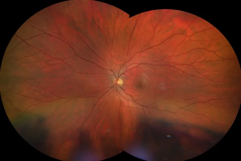
Edema
Fluid and fats seep out of damaged vessels into the retina. If this affects the macula, macular edema develops and vision becomes blurry.
Ischemia
Closed vessels leave areas of the retina short of oxygen. These send out growth signals (VEGF).
Proliferation
New vessels grow onto the retina and into the vitreous. They bleed easily and can form scars that pull on the retina.
Duration, sugar, blood pressure.
The longer the diabetes has lasted and the higher blood sugar and blood pressure are over the years, the more likely it is. With type 1 diabetes, after 20 years almost everyone shows at least mild changes; with type 2, about one in ten, and after more than ten years of diabetes about one in four, has retinopathy, some already at diagnosis.
The strongest factor. That is why in type 2 the follow-up checks begin immediately, in type 1 after a few years.
Every percentage point lower reduces the risk significantly. What matters are the values over the years, not a single value.
Puts additional strain on the vessels. Well-controlled blood pressure protects the eyes and kidneys.
Protein in the urine and high blood fats go along with more retinal damage.
Hormonal changes can speed up retinopathy.
Damages the vessels further and worsens the supply to the retina.
Not at all for a long time. That is the insidious part.
In the early stages you see completely normally. Symptoms only develop when the macula swells or vessels bleed. Many people with retinopathy that needs treatment still have full visual acuity. A sudden worsening, on the other hand, is an emergency.
Only the eye doctor can see small bulges and bleeding. That is why the follow-up checks matter, even if everything seems fine.
Reading becomes hard, colors look paler, straight lines look slightly distorted. Develops over weeks to months.
Sometimes better, sometimes worse, often along with blood sugar values. It can also be due to the lens; the examination clarifies it.
Many black dots, a red or dark veil: signs of bleeding into the vitreous. Report it the same day.
May be a retinal detachment caused by scar traction. Have your eyes examined by an eye doctor immediately.
Diabetes also makes a cataract and glaucoma more likely. We look at these as well at every follow-up check.
From invisible to threatening.
Doctors distinguish non-proliferative retinopathy (mild, moderate, severe) from proliferative retinopathy, in which new vessels grow. Independently of this, macular edema can occur at any stage. Without treatment, about every second person with severe non-proliferative retinopathy develops proliferative retinopathy with new vessels within a year.
Tap a stage.
Normal retinal vessels.
Nothing.
Follow-up check every 1–2 years depending on general risk. Keep sugar and blood pressure on target.
A few microaneurysms, small bulges of the vessel wall.
Nothing.
Follow-up check usually annually. No eye treatment needed, but improve general values.
More bleeding, hard exudates, first signs of reduced blood flow.
Usually nothing, except with macular edema.
Follow-up check about every 6 months; look for macular edema.
Many hemorrhages in all quadrants, beaded veins, vessel abnormalities (4-2-1 rule).
Often still nothing.
Follow-up check every 3 months; high risk of new vessels, consider laser or injection.
New vessels on the optic nerve or retina, bleeding into the vitreous, scars.
With bleeding, a sudden veil or soot rain.
Treatment: laser of the peripheral retina and/or injections; with bleeding or traction, surgery.
Thickened, fluid-filled macula on OCT.
Blurry vision in the center, reading is hard.
When the center is involved, usually injections; follow-up with OCT.
| Stage | Findings | Follow-up check | Treatment of the eye |
|---|---|---|---|
| No retinopathy | normal | 1–2 years | none |
| Mild non-proliferative | Microaneurysms | yearly | none |
| Moderate non-proliferative | Hemorrhages, exudates | 6 months | usually none |
| Severe non-proliferative | 4-2-1 rule met | 3 months | Consider laser or injection |
| Proliferative | new vessels | according to plan, close-meshed | Laser, injection, surgery if needed |
| Macular edema (center affected) | Thickening on OCT | during therapy 4–16 weeks | Injection, laser if needed |
Classification according to the international classification (ETDRS/AAO), simplified.
One hour for your retina.
The eye doctor checks visual acuity and eye pressure and looks at the entire retina, classically with pupil-dilating drops. At dr.vision, the retinal image can often be taken without drops using the ultrawide image; if the pupils are dilated after all, you must not drive yourself for a few hours afterward. An OCT scan shows whether the macula is swollen.

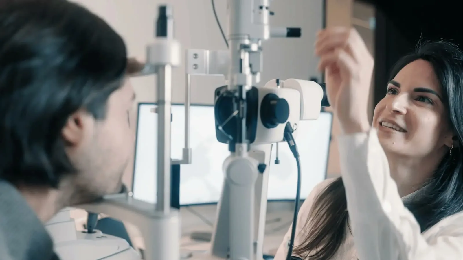
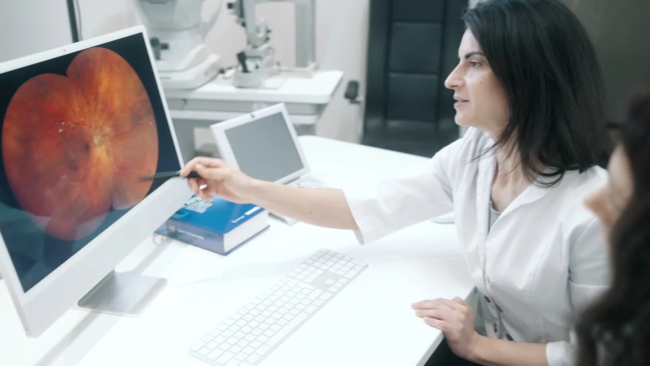
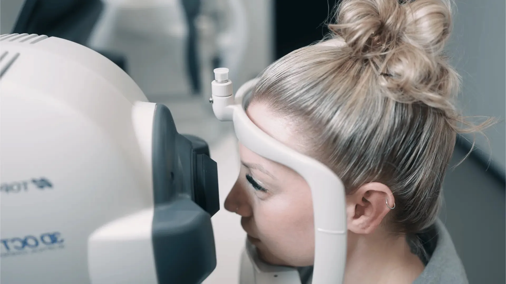
 Step 1 · Conversation
We ask about the type of diabetes, HbA1c, blood pressure and your medications. Bring the notification form from your family doctor or your latest lab values.
Step 2 · Vision test and pressureWith your glasses for distance and near. Plus the eye pressure and a look at the lens, because diabetes also makes cataract and glaucoma more likely.
Step 3 · Retinal imageWith the ultrawide image we see a large part of the retina, often without dilating the pupils. The image is saved and compared next time.
Step 4 · OCTThe cross-sectional scan measures the thickness of the macula to thousandths of a millimeter. It finds macular edema before you notice it.
Step 5 · FindingsYou learn right away whether and which changes are present and when your next follow-up check is due. The findings go to your family doctor or diabetes specialist.
All examinations are contact-free or with brief numbing. Including the conversation, around 45–60 minutes.
Then your vision is blurry and sensitive to glare for a few hours. Please do not drive yourself, and bring sunglasses.
Fluorescein angiography (dye into the arm vein) or OCT angiography show closed vessels and new vessels more precisely.
Water in the center of vision.
In diabetic macular edema, fluid collects in the macula, the spot of sharpest vision. It is the most common cause of worse vision in diabetes and can occur at any stage of retinopathy. If the center is affected and vision is reduced, injections into the eye are the standard treatment; many patients regain visual acuity with them.
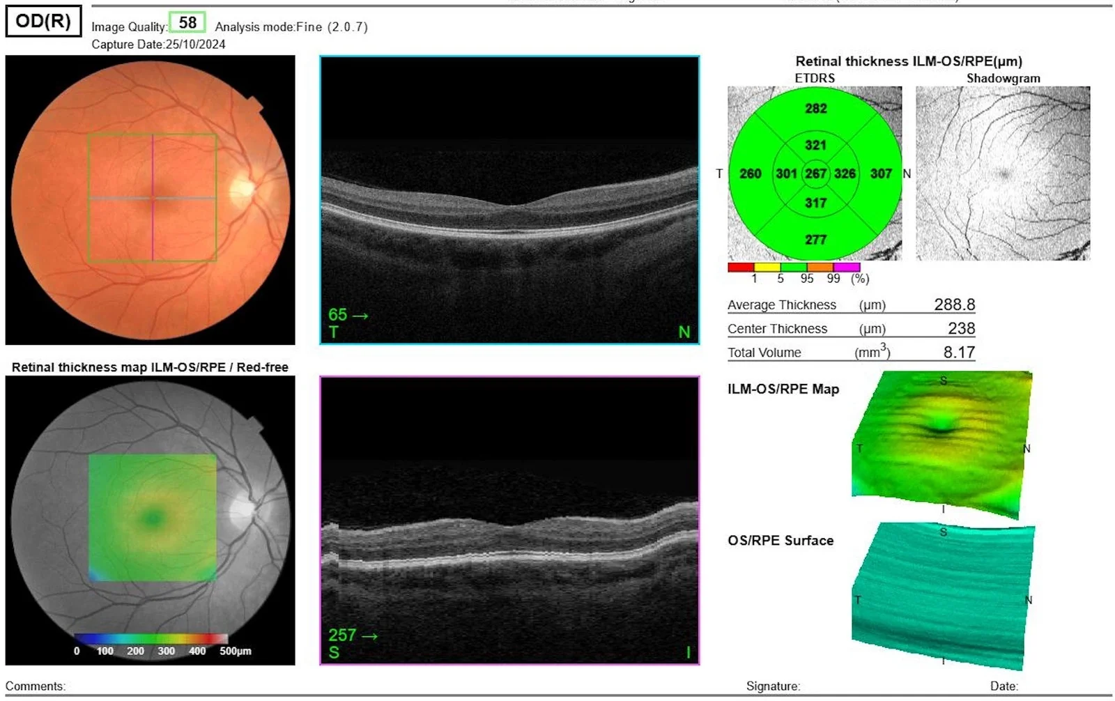
OCT
Whether the macula is thickened and whether the center is affected is reliably shown only by OCT. The treatment depends on it.
Anti-VEGF
At the start usually several injections four weeks apart, then less often depending on the findings. If the response is insufficient, cortisone implants are an option.
1–3 years
With macular edema, the intervals often become long over time; some patients no longer need injections after a few years.
Foundation, injection, laser, surgery.
The foundation is always general control: blood sugar, blood pressure and blood fats on target. For the eye there are three paths: injections for macular edema and as an option for new vessels, laser for proliferative retinopathy, and surgery for vitreous bleeding or traction on the retina. Treated in time, severe vision loss can be prevented in most cases.
| Criterion | Injection (anti-VEGF, IVOM) | Laser (panretinal / focal) | Surgery (vitrectomy) |
|---|---|---|---|
| Used for | Macular edema; proliferative retinopathy as an alternative or addition | proliferative and severe non-proliferative retinopathy; selected edemas | Vitreous bleeding that does not clear; traction on or detachment of the retina |
| Effect | reduces edema, makes new vessels regress | ablates poorly supplied retina, lowers VEGF lastingly | removes blood and scar tissue, reattaches the retina |
| Visual acuity | often improves with edema | preserves; may somewhat limit night vision and visual field | depends on the starting point |
| How often | Start with several injections, then every 4–16 weeks | usually 2–4 sessions | once, rarely repeated |
| Duration of effect | weeks, hence repeated | usually long-lasting | lasting |
| Risks | red spot common; infection about 1 in 1,800–2,000 injections | limited night and peripheral vision; rarely temporarily more edema | cataract common afterward; rarely retinal detachment or infection |
| Burden | brief, numbing drops | sessions of minutes, sometimes uncomfortable | surgery with anesthesia, recovery takes weeks |
First choice for macular edema.
If the center of the retina is affected in diabetic macular edema and vision is worse, anti-VEGF injections are the standard treatment. In studies, patients regained on average about 2 lines of visual acuity. In proliferative retinopathy, too, injections make new vessels regress, but they work only as long as treatment continues regularly.
Numbing drops, disinfection, a fine needle, a few seconds. You are in the practice for about an hour. Prof. Dr. Mehdi Shajari performs the injection personally.
Anti-VEGF drugs such as ranibizumab, aflibercept or faricimab; if the response is insufficient, a cortisone implant.
Step by step, fear and pain, treatment schedules, risks and costs: on the page Injection therapy (IVOM).
When new vessels grow.
In proliferative retinopathy, panretinal laser coagulation ablates the poorly supplied peripheral retina. This lowers the growth signal, and new vessels regress. The laser lowers the risk of severe vision loss in high-risk proliferative retinopathy by more than half, and the effect usually lasts a long time.
Hundreds to thousands of small laser spots at the edge of the retina, usually in 2–4 sessions.
2–4sessions
Targeted spots at leaking sites outside the center. Today mostly a supplement to injections.
Planning on the retinal image, automatically placed spots, often gentler.
When blood does not clear or scars pull.
If a vitreous bleed does not clear on its own after a few weeks, or if scar tissue lifts the retina, the vitreous is removed surgically (vitrectomy). The blood is removed, scars are released and the retina is treated with laser. Thanks to earlier treatment, surgery is needed much less often today.
Sudden dark veil. Often we wait a few weeks to see whether the blood clears; with type 1 diabetes or detachment, surgery is done earlier.
Scars pull the retina away. If this threatens the macula, surgery is done promptly.
Retinal surgery is performed at specialized centers. We examine, advise and refer you.
Before, during, after.
With existing type 1 or type 2 diabetes, retinopathy can progress faster during pregnancy. That is why: a retinal follow-up check when you want to have a child or, at the latest, when the pregnancy is confirmed, then about every 3 months until birth, more often if there are changes. Gestational diabetes alone does not increase the retinal risk.
| Time | What we recommend | Why |
|---|---|---|
| Wanting to have a child | Retinal follow-up check, optimize values | treat retinopathy that needs treatment beforehand |
| Pregnancy confirmed | Follow-up check as soon as possible | Baseline findings for comparison |
| During pregnancy | every 3 months, more often with retinopathy | Hormones, blood volume and rapid lowering of sugar can speed it up |
| After birth | Follow-up check in the first 3–6 months | Worsening partly regresses, partly does not |
| Gestational diabetes only | no additional retinal follow-up check (NVL) | no increased retinal risk; diabetes check after birth with the family doctor |
The drops for the examination are used during pregnancy after weighing the pros and cons, because the follow-up check is important. The retinal image without drops is an alternative.
Anti-VEGF drugs are avoided during pregnancy where possible. If treatment is needed, we discuss alternatives such as laser together with your gynecologist.
Retinopathy is only rarely a reason against a natural birth. If needed, we coordinate with the maternity hospital.

Follow-up check due?
Call us. We will take a look.
Jana from our Patient Care arranges your retinal follow-up check, at off-peak hours if you wish. Bring the notification form from your family doctor or your latest values. Reachable around the clock.
Your path. In four steps.
From booking the appointment to the findings for your practice: the follow-up check takes about an hour, and you learn the result the same day.
Step 1 of 4
Bring your values, ask questions.
Patient Care tells you in advance whether your pupils will be dilated and you should not drive yourself.
Arrange an appointmentStep 2 of 4
Retina, macula, lens, pressure.
The images are saved and compared at the next follow-up check.
The examination in detailStep 3 of 4
Result and schedule.
If treatment is needed, you receive a written plan and cost estimate.
The stagesStep 4 of 4
Stay on track.
Patient Care reminds you of every appointment.
Your scheduleKeep sugar and blood pressure well controlled. Keep your follow-up checks.
Good blood sugar and blood pressure values over the years are the most effective protection. In large studies, better control reduced the risk of retinopathy by about a fifth to three quarters. It is also important to keep your follow-up checks, even if you see well.
Your diabetes specialist sets your personal target. Every lasting reduction protects the retina.
Protects retina, kidneys and heart. Tell us your values.
If sugar is lowered sharply within a short time, for example with new medications or an insulin pump, existing retinopathy can temporarily worsen. Then have closer follow-up checks.
Smoking damages the vessels further, in the eyes, heart and legs.
Allowed as long as visual acuity and visual field meet the requirements. After extensive laser, we check the visual field.
Schedule the eye follow-up check next to your annual diabetes check. Patient Care reminds you.

Because your retina should not wait.
At dr.vision in Frankfurt: retinal diagnostics often without pupil dilation, physicians with a focus on the retina, and findings that go to your practice. 5.0 stars from 376 reviews.
Three specialist physicians. One team.
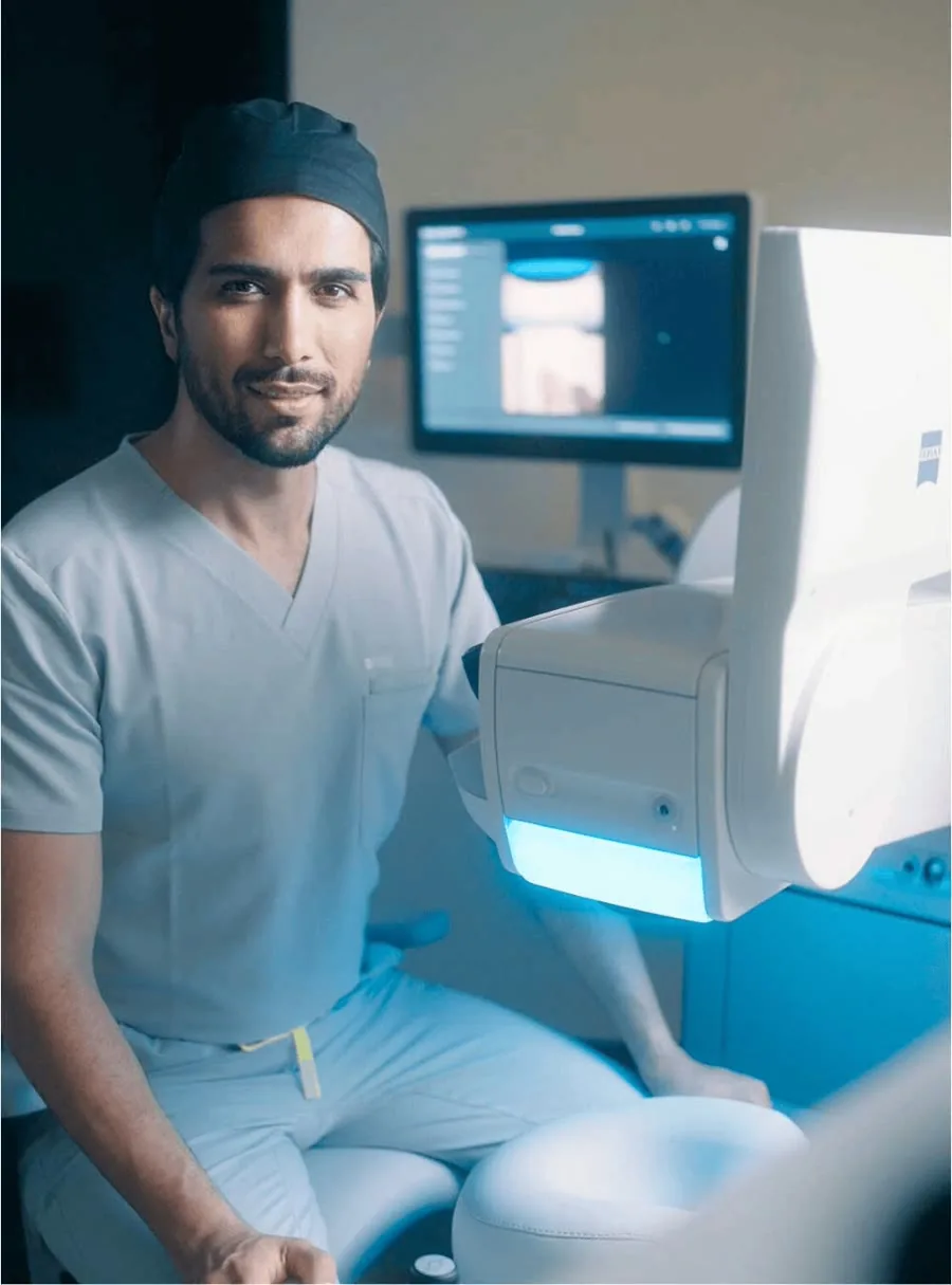
Dr. Claudia Grunwald has her areas of focus in, among other things, the diagnostics and treatment of retinal diseases; Ivana Pavlovic was a senior physician (Oberärztin) at the University Hospital Frankfurt. Prof. Dr. Mehdi Shajari, specialist physician and professor of ophthalmology (German title: außerplanmäßiger Professor), performs injection therapies personally. You are looked after not by changing doctors, but by the same team.
What you get with us.
One team
Dr. Grunwald, senior physician Pavlovic, Prof. Dr. Mehdi Shajari.
Reviews
5,0
376 Google reviews
Without drops
Afterward you can usually drive home yourself.
Ultrawide retinal image, OCT of the macula, vision test and eye pressure in one appointment. You go home with findings and a schedule.
5,0
★★★★★
from 376 Google reviews
Whoever knows your retina sees changes sooner.
Image
Every image compared with the last.
Family doctor or diabetes specialist receives the result.
Values, stage, pregnancy: we take the time until everything is clear.
Monday to Friday until 19:00, Saturday until 13:00.
Billing per the German fee schedule for physicians (GOÄ), written cost estimate before every treatment.
Jana answers questions about appointments, findings and costs via WhatsApp, usually within 10–15 minutes, Mon–Fri 8:00–19:00, Sat 9:00–15:00. We are available by phone around the clock.
Patient Care
Junghofstraße 16, free parking in the building.
A look inside the practice and the team.



Transparent. In writing, in advance.
The follow-up check is billed as a private medical service per the German fee schedule for physicians (GOÄ), depending on scope with retinal image and OCT of the macula. Private health insurance and Beihilfe (allowance for German civil servants) often cover a large part of it depending on the plan, because it is medically necessary in diabetes; you should plan for a share of your own. Patients with statutory health insurance pay with us themselves.
Per the German fee schedule for physicians (GOÄ)Costs depend on scope · cost estimate before every treatment
The retinal follow-up check for diabetes is medically necessary; private health insurers often cover a large part of it depending on the plan, as well as OCT and treatments, though a share of your own usually remains. Before every treatment you receive a cost estimate.
The medically necessary services per the German fee schedule for physicians (GOÄ) are eligible for Beihilfe (allowance for German civil servants). Your private supplementary insurance reimburses the remaining share according to its plan; you should plan for a share of your own.
You pay for the follow-up check with us yourself. Billing is according to the German fee schedule for physicians (GOÄ), and we tell you the costs in advance. Costs you bear yourself can be declared in your tax return as extraordinary expenses. Whether and how much the tax office recognizes depends, among other things, on your reasonable burden. More under Tax.
| Follow-up schedule | without retinopathy: every 2 years with a low, annually with an elevated general risk; with retinopathy according to the eye doctor's plan |
|---|---|
| First follow-up check | Type 2 at diagnosis; type 1 from 5 years of diabetes or from about age 11 |
| Pregnancy | at planning/confirmation, then about every 3 months; gestational diabetes alone without additional follow-up check |
| Disease | Damage to the retinal vessels from diabetes; long without symptoms |
| Stages | mild, moderate, severe non-proliferative; proliferative; macular edema at any stage |
| Diagnostics | Vision test, eye pressure, ultrawide retinal image often without pupil dilation, OCT of the macula |
| Treatment | Foundation: sugar and blood pressure; injections for macular edema; laser for proliferative retinopathy; vitrectomy for bleeding or traction |
| Emergency | sudden veil, soot rain, curtain: the same day |
| Costs | under the GOÄ |
| Insurance | private health insurance and Beihilfe per the GOÄ, patients with statutory health insurance are self-paying patients at dr.vision |
| Doctors | Dr. Claudia Grunwald, senior physician Ivana Pavlovic, Prof. Dr. Mehdi Shajari |
| Location | Junghofstraße 16, 60311 Frankfurt am Main |
Go deeper.
This page answers the most important questions. For related topics there are separate pages, each with context and the questions from consultation hours:
Medications, process, fear and pain, treat-and-extend.
To the IVOM pageMaculaThe other common disease of the center of the retina, with the Amsler test.
To the AMD pagePreventive careRetina, optic nerve and lens in one examination.
To the check-upLensMore common and earlier with diabetes: when to operate?
To the cataract pageOptic nerveDiabetes increases the risk, also for special forms.
To the glaucoma pageVitreousSpots in your field of view: when harmless, when bleeding or an emergency.
Learn moreStill have questions?
The questions we hear most often in consultation hours, with our answers. Prof. Dr. Mehdi Shajari
Every two years with no retinal changes if your general risk is low, otherwise annually. If retinopathy or macular edema is already known, the eye doctor sets shorter intervals, often every three to six months. If your vision suddenly worsens, come immediately.
With type 2 diabetes, immediately at diagnosis, because the diabetes has often been present for years. With type 1 diabetes, from about five years of diabetes, in children from about eleven years. After that, at the schedule your eye doctor sets based on findings and general risk.
Because diabetic retinopathy causes no symptoms for a long time. Even changes that need treatment and an early macular edema can be present with full visual acuity. Anyone who comes only when vision worsens often already has damage that can no longer be fully reversed.
If the pupils are not dilated, yes. With the ultrawide image, the retinal image is often possible without drops. If the pupils are dilated after all, your vision is blurry for a few hours and you must not drive yourself. Patient Care tells you beforehand what to expect.
Untreated, diabetic retinopathy can lead to severe vision loss; in people of working age it is one of the most common causes. With regular follow-up checks and timely treatment, severe vision loss can be prevented in most cases.
Early changes can partly regress with better control; dead retina does not recover. A macular edema can often be pushed back significantly with injections. New vessels regress after laser or injections, scars remain.
With existing retinopathy, a very rapid, strong lowering of blood sugar can temporarily lead to a worsening. That is no reason not to improve your treatment, but it is a reason for a retinal follow-up check beforehand and close follow-up checks in the months afterward. Tell us when your therapy is changed.
That depends on the findings. In macular edema involving the center of the retina, injections are the first choice. In proliferative retinopathy, laser and injections both work; the laser usually works long-term, sometimes a follow-up treatment is needed; the injection has to be repeated regularly. Often both are combined.
The surface of the eye is numbed with drops. With laser at the edge of the retina, many people feel a stinging or pulling, especially in the peripheral areas. The treatment is therefore spread over several short sessions. Afterward you will see glare for a few hours.
According to the National Disease Management Guideline, no additional retinal follow-up check is needed with gestational diabetes alone, because it does not increase the retinal risk. It is different with pre-existing type 1 or type 2 diabetes: then a follow-up check at the start and about every three months until birth.
With existing diabetes, retinopathy can progress faster during pregnancy, especially if it was already present beforehand or if blood sugar is lowered quickly at the start. That is why the retina is checked closely before and during pregnancy. Many changes regress after birth.
Patients with statutory health insurance pay for the follow-up check with us themselves. Billing is according to the German fee schedule for physicians (GOÄ), and we tell you the costs in advance. Private insurers and Beihilfe (allowance for German civil servants) often cover a large part of the follow-up check depending on the plan; you should plan for a share of your own.
Billing is according to the German fee schedule for physicians (GOÄ), depending on which examinations are needed, such as retinal image and OCT. You receive an estimate of the costs in advance and a written cost estimate for every treatment. The report goes to your family doctor or diabetes specialist.
Medically reviewed by Prof. Dr. Mehdi Shajari · As of October 2026
Everything important to read again and pass on.
Arrives by e-mail in about 60 seconds. No newsletter, just the guide and, if you wish, a follow-up from Patient Care. Privacy policy
How would you like to continue?
The follow-up check includes a vision test, eye pressure, a retinal image, an OCT if needed, and a conversation about the findings and schedule. You go home with a clear result and your follow-up schedule, and your practice receives a report.
In Frankfurt, Junghofstraße 16 · with retinal image and report to your practice
Our locations.
 Opening hours
Junghofstraße 16, 60311 Frankfurt am Main
 Opening 2027
Breite Straße 69, 40213 Düsseldorf (in the Neuer Stahlhof, corner of Graf-Adolf-Platz)
Until opening: preliminary talk in Frankfurt, Patient Care plans your travel.
Plan route Opening 2027
Lautenschlagerstraße 23, 70173 Stuttgart (2nd floor, step-free access)
Until opening: preliminary talk in Frankfurt, Patient Care plans your travel.
Plan route